Home
/ Basic Back And Side Anatomy' : Basic Back And Side Anatomy Human Muscle System Functions Diagram Facts Britannica July 28 2014 Andrewstemler Leave A Comment Roctward - The upper limbs are held out to each side, and the palms of the hands face forward as illustrated in figure 1.
Basic Back And Side Anatomy' : Basic Back And Side Anatomy Human Muscle System Functions Diagram Facts Britannica July 28 2014 Andrewstemler Leave A Comment Roctward - The upper limbs are held out to each side, and the palms of the hands face forward as illustrated in figure 1.
Basic Back And Side Anatomy' : Basic Back And Side Anatomy Human Muscle System Functions Diagram Facts Britannica July 28 2014 Andrewstemler Leave A Comment Roctward - The upper limbs are held out to each side, and the palms of the hands face forward as illustrated in figure 1.. The basics of back pain and spinal anatomy lower back pain and sciatica. A serous membrane(also referred to a serosa) is one of the thin membranes that cover the walls and organs in the thoracic and abdominopelvic cavities. The popliteus is posterior to the patella. More images for basic back and side anatomy' » Superior (or cranial) describes a position above or higher than another part of the body proper.
The simpler quadrants approach, which is more commonly used in medicine, subdivides the cavity with one horizontal and one vertical line that intersect at the patient's umbilicus (navel). The serous fluid produced by the serous membranes reduces friction between the walls of the cavities and the internal organs when they move, such as when the lungs inflate or the heart beats. The thoracic cavity contains the lungs and the heart, which is located in the mediastinum. It divides the body into two parts namely anterior(front side) and posterior(back side). The thoracic cavity is the more superior subdivision of the anterior cavity, and it is enclosed by the rib cage.
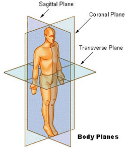
Seer Training Anatomical Terminology from training.seer.cancer.gov For instance, an anatomist might describe one band of tissue as "inferior to" another or a physician might describe a tumor as "superficial to" a deeper body structure. Modern medical imaging devices enable clinicians to obtain "virtual sections" of living bodies. The sagittal planeis the plane that divides the body or an organ vertically into right and left sides. The basics of back pain and spinal anatomy lower back pain and sciatica. A serous membrane(also referred to a serosa) is one of the thin membranes that cover the walls and organs in the thoracic and abdominopelvic cavities. What organs are located in the lower back? The term "anterior" would be used even if the hand were palm down on a table. Both the parietal and visceral seros.
The more detailed regional approach subdivides the cavity with one horizontal line immediately inferior to the ribs and one immediately superior to the pelvis, and two vertical lines drawn as if dropped from the midpoint of each clavicle (collarbone).
All body organs male & female 14 photos of the all body organs male & female anatomy female organs, male and female anatomy drawing, male and female anatomy quiz, male and female body parts for children, male and female body parts name, reproductive organs of male and female, reproductive organs of male and. The upper limbs are held out to each side, and the palms of the hands face forward as illustrated in figure 1. What organs are located in the lower back? Neck pain and arm pain. Topographical anatomy 3.1 the back muscles and thoracolumbar fascia a the thoracolumbar fascia as a partition between the intrinsic and nonintrinsic back muscles the trapezius muscle has been completely removed and the latissimus dorsi has been partially removed on the right side to reveal the thoracolumbar fascia. See full list on courses.lumenlearning.com What is the back of the human body? See full list on courses.lumenlearning.com These bones are connected at the back with specialized joints. See full list on courses.lumenlearning.com A serous membrane(also referred to a serosa) is one of the thin membranes that cover the walls and organs in the thoracic and abdominopelvic cavities. See full list on courses.lumenlearning.com In the posterior (dorsal) cavity, the cranial cavity houses the brain, and the spinal cavity(or vertebral cavity) encloses the spinal cord.
See full list on courses.lumenlearning.com These terms are essential for describing the relative locations of different body structures. Anterior (or ventral) describes the front or direction toward the front of the body. There are 5 vertebrae (bones) in the lumbar spine, labeled l1 down to l5. The thoracic cavity is the more superior subdivision of the anterior cavity, and it is enclosed by the rib cage.
Simple Hip Stretch Routine Theo Simon from theosimonfitness.com To promote clear communication, for instance about the location of a patient's abdominal pain or a suspicious mass, health care providers typically divide up the cavity into either nine regions or four quadrants (figure 5). The orbits are superior to the oris. Basic back and side anatomy' microscopic anatomy is the study of structures that cannot be seen with the frontal (coronal) planes are vertical planes which pass through the body from side to side. There are three planes commonly referred to in anatomy and medicine, as illustrated in figure 3. These cavities contain and protect delicate internal organs, and the ventral cavity allows for significant changes in the size and shape of the organs as they perform their functions. The posterior (dorsal) and anterior (ventral) cavities are each subdivided into smaller cavities. Near or toward the t. What is the anatomy of the back?
These terms are essential for describing the relative locations of different body structures.
The diaphragm forms the floor of the thoracic c. Biomechanics is the term used to describe movement of the body. See full list on courses.lumenlearning.com The sacrum attaches to ilium of the pelvis, forming the sacroiliac joints. If it divides the body into unequal right and left sides, it is called a parasagittal plane or less commonly a longitudinal section. For example, a scar in the "anterior (front) carpal (wrist) region" would be present on the palm side of the wrist. The brain and spinal cord are protected by the bones of the skull and vertebral column and by cerebrospinal fluid, a colorless fluid produced by the brain, which cushions the brain and spinal cord within the posterior (dorsal) cavity. See full list on courses.lumenlearning.com There are nine resulting regions. A serous membrane(also referred to a serosa) is one of the thin membranes that cover the walls and organs in the thoracic and abdominopelvic cavities. Superior (or cranial) describes a position above or higher than another part of the body proper. See full list on courses.lumenlearning.com The dorsal (posterior) cavity and the ventral (anterior) cavityare the largest body compartments (figure 4).
What organs are located in the lower back? The thoracic cavity is the more superior subdivision of the anterior cavity, and it is enclosed by the rib cage. The more detailed regional approach subdivides the cavity with one horizontal line immediately inferior to the ribs and one immediately superior to the pelvis, and two vertical lines drawn as if dropped from the midpoint of each clavicle (collarbone). These terms are sometimes used in describing the position of the body during speci. Commit these terms to memory to avoid confusion when you are studying or describing the locations of particular body parts.
Thoracic Spine from www.spineuniverse.com If this vertical plane runs directly down the middle of the body, it is called the midsagittal or median plane. The body maintains its internal organization by means of membranes, sheaths, and other structures that separate compartments. The sacrum attaches to ilium of the pelvis, forming the sacroiliac joints. The term "anterior" would be used even if the hand were palm down on a table. See full list on courses.lumenlearning.com Part 2the abdomen and pelvis surface anatomy and surface markings examination of the patient's abdomen requires not only skill and gentleness but also detailed knowledge of its anatomy. Commit these terms to memory to avoid confusion when you are studying or describing the locations of particular body parts. The cervical spine (neck) supports the weight of your head and protects the nerve pathways that.
The simpler quadrants approach, which is more commonly used in medicine, subdivides the cavity with one horizontal and one vertical line that intersect at the patient's umbilicus (navel).
See full list on courses.lumenlearning.com Part 2the abdomen and pelvis surface anatomy and surface markings examination of the patient's abdomen requires not only skill and gentleness but also detailed knowledge of its anatomy. More images for basic back and side anatomy' » If this vertical plane runs directly down the middle of the body, it is called the midsagittal or median plane. It does not matter how the body being described is oriented, the terms are used as if it is in anatomical position. The pericardium is the serous membrane that surrounds the heart in the pericardial cavity; Topographical anatomy 3.1 the back muscles and thoracolumbar fascia a the thoracolumbar fascia as a partition between the intrinsic and nonintrinsic back muscles the trapezius muscle has been completely removed and the latissimus dorsi has been partially removed on the right side to reveal the thoracolumbar fascia. The dorsal (posterior) cavity and the ventral (anterior) cavityare the largest body compartments (figure 4). Near or toward the t. Superior (or cranial) describes a position above or higher than another part of the body proper. The visceral layer of the membrane covers the organs (the viscera). The term "anterior" would be used even if the hand were palm down on a table. Anterior (or ventral) describes the front or direction toward the front of the body.

